Your heart is a muscular organ at the centre of your circulatory system. It is needed for pumping blood around the body, but how does blood travel around? Thankfully, you have blood vessels for this. Arteries and veins are tube-shaped vessels - blood leaves the heart in arteries and returns through veins.
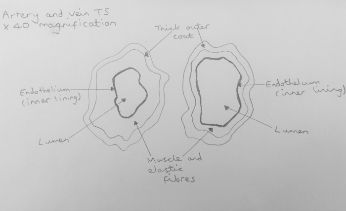
Arteries and veins have layers to their structure but the relative thickness of these layers is different. Both types of blood vessel have a tough, outer layer. Arteries carry blood at high pressure, so they have thick, muscular walls (thin walls would easily break under the high pressure at which the heart pumps blood). The lumen (space inside the middle of the tube) is narrow to maintain the high pressure. Veins return blood to the heart at low pressure, so their walls are considerably thinner compared to arteries and have less muscular tissue. The lumen is much wider.
In this worksheet, you will learn how a light microscope is used to see a transverse section of an artery and a vein. A transverse section is sometimes called a cross-section. Imagine cutting something like a cake in half - you will be able to see the inside, this is the cross-section. Close attention needs to be paid to the features seen inside both blood vessels, as detailed scientific diagrams need to be drawn based on these careful observations.
Method:
1. You are given a pre-prepared slide of a transverse section (T.S.) of an artery and a vein.
2. Set the objective lens of the microscope to a low magnification (x4) and examine the T.S. of the artery closely. You may need to switch to a x40 magnification to see the layers of the artery in more detail.
3. Very carefully, draw a labelled diagram of the artery, making sure you include the tough, outer layer, the muscular and elastic fibres, the endothelium and the lumen. It is very important to capture the relative thickness of the different layers in your diagram, as you need to be able to make comparisons with the vein. Use a pencil to draw smooth, continuous lines (do not leave gaps). This is science, not art, so you do not need to include shading or colour - it's a diagram, not a picture.
4. Repeat the steps above but this time for the vein slide.

The total magnification of the image seen under the microscope can be worked out by multiplying the power of the objective lens (x 4) by the power of the eyepiece lens (x 10).
You may also be asked to work out the magnification of a blood vessel in your diagram compared to the real blood vessel on the slide. All they are asking you is how much larger is the blood vessel in your diagram compared to the real thing.
Example:
The diameter of the artery on the slide is 4 mm.
Draw a line across the diagram of the artery to mark the diameter.
The length of this line needs to be measured with a ruler - the diameter is 68 mm in this example.
Use the equation:
magnification = image size ÷ actual size
magnification = 68 ÷ 4
magnification = 17
This shows that your diagram of the artery was drawn 17 times larger than the real artery actually is.
Are you ready to have a go at some questions now?


.PNG)


